Breast MRI
A Case-Based Approach
Nonfiction, Health & Well Being, Medical, Specialties, Radiology & Nuclear Medicine| Author: | Caren Greenstein | ISBN: | 9781451154306 |
| Publisher: | Wolters Kluwer Health | Publication: | March 29, 2012 |
| Imprint: | LWW | Language: | English |
| Author: | Caren Greenstein |
| ISBN: | 9781451154306 |
| Publisher: | Wolters Kluwer Health |
| Publication: | March 29, 2012 |
| Imprint: | LWW |
| Language: | English |
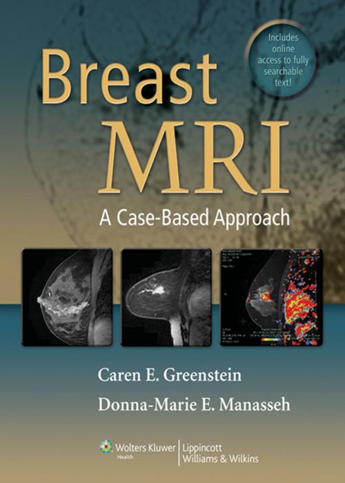
MRI is increasingly being used by radiologists to confirm diagnoses and perform operative procedures of the breast. MRI's contrast between soft tissues in the breast is many times greater than that obtained by plain-film mammography. As opposed to x-rays, which are known to cause damage to cellular DNA, the magnetic fields and radiowaves used with MRI are not known to have any long-term biologic effect. MRI of the breast requires intravenous injection of a contrast agent, which helps highlight breast abnormalities. The American Cancer Society has advised women at high risk for breast cancer to have an MRI. This book, a collaboration between an experienced breast imager and a breast surgeon, contains 100 cases and covers high risk screening, extent of disease evaluation, as well as the full range of benign and malignant tumors found in the breast: DCIS, invasive ductal cancers, and invasive lobule cancers. Rare lesions such as phyllodes, mucinous, liposarcomas, and myotiboblastomas are also covered. Pathologic correlations are included where appropriate. This book has a special emphasis on preoperative planning involving MRI and thus may also appeal to surgical oncologists specializing in breast cancer.
MRI is increasingly being used by radiologists to confirm diagnoses and perform operative procedures of the breast. MRI's contrast between soft tissues in the breast is many times greater than that obtained by plain-film mammography. As opposed to x-rays, which are known to cause damage to cellular DNA, the magnetic fields and radiowaves used with MRI are not known to have any long-term biologic effect. MRI of the breast requires intravenous injection of a contrast agent, which helps highlight breast abnormalities. The American Cancer Society has advised women at high risk for breast cancer to have an MRI. This book, a collaboration between an experienced breast imager and a breast surgeon, contains 100 cases and covers high risk screening, extent of disease evaluation, as well as the full range of benign and malignant tumors found in the breast: DCIS, invasive ductal cancers, and invasive lobule cancers. Rare lesions such as phyllodes, mucinous, liposarcomas, and myotiboblastomas are also covered. Pathologic correlations are included where appropriate. This book has a special emphasis on preoperative planning involving MRI and thus may also appeal to surgical oncologists specializing in breast cancer.


