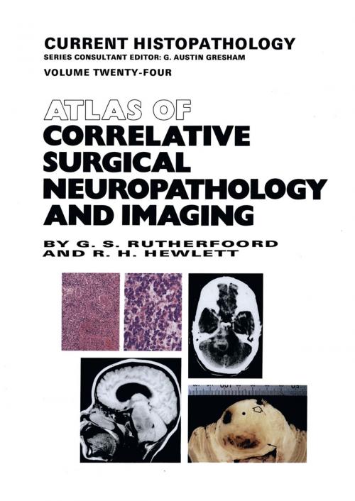
Atlas of Correlative Surgical Neuropathology and Imaging
Nonfiction, Health & Well Being, Medical, Specialties, Pathology, Internal Medicine, Neurology| Author: | G.S. Rutherfoord, R.H. Hewlett | ISBN: | 9789401114349 |
| Publisher: | Springer Netherlands | Publication: | December 6, 2012 |
| Imprint: | Springer | Language: | English |
| Author: | G.S. Rutherfoord, R.H. Hewlett |
| ISBN: | 9789401114349 |
| Publisher: | Springer Netherlands |
| Publication: | December 6, 2012 |
| Imprint: | Springer |
| Language: | English |
Because of the topographic and pathophysiologic information obtained with contemporary neuroimaging techniques, CT and MR scanning now constitute the most important investigation in clinical neurology. In many instances of mass lesions, the images provide a reliable or near-definitive diagnosis, and make possible the accurate and even selective acquisition of biopsy samples.
For pathologists and neuropathologists rendering a brain biopsy service, a basic knowledge of CT and MR scanning is now mandatory, and the objective of this atlas is to present the principles of neuroimaging through clinicopathological correlation.
It contains a wide range of clinical material, with over 600 CT and MR images correlated with over 400 full-colour pathomorphological micrographs. A full discussion of differential diagnosis is complemented by extensive references.
Although aimed mainly at pathologists in neurosurgical practice, the atlas will also benefit neurosurgeons and radiologists, especially those in training.
