Anterior Segment Optical Coherence Tomography
Nonfiction, Health & Well Being, Medical, Specialties, Ophthalmology| Author: | ISBN: | 9781617112089 | |
| Publisher: | SLACK Incorporated | Publication: | January 15, 2008 |
| Imprint: | SLACK Incorporated | Language: | English |
| Author: | |
| ISBN: | 9781617112089 |
| Publisher: | SLACK Incorporated |
| Publication: | January 15, 2008 |
| Imprint: | SLACK Incorporated |
| Language: | English |
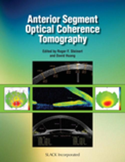
High-speed anterior segment optical coherence tomography (OCT) offers a non-contact method for high resolution cross-sectional and three-dimensional imaging of the cornea and the anterior segment of the eye. As the first text completely devoted to this topic, Anterior Segment Optical Coherence Tomography comprehensively explains both the scientific principles and the clinical applications of this exciting and advancing technology. Anterior Segment Optical Coherence Tomography enhances surgical planning and postoperative care for a variety of anterior segment applications by expertly explaining how abnormalities in the anterior chamber angle, cornea, iris, and lens can be identified and evaluated using the Visante OCT™. Inside Anterior Segment Optical Coherence Tomography, Dr. Roger Steinert and Dr. David Huang, along with 22 of the field's leading professionals, provide a wealth of useful clinical and physiological material about this new diagnostic imaging technique. Valuable images are included to assist in the pre- and postoperative assessment of various anterior segment disorders. On the leading edge of anterior segment imaging: Mapping of corneal thickness and keratoconus evaluation Measurement of LASIK flap and stromal bed thickness Visualization and measurement of anterior chamber angle and diagnosis of narrow angle glaucoma Measuring the dimensions of the anterior chamber and assessing the fit of intraocular lens implants Visualizing and measuring the results of corneal implants and lamellar procedures Imaging through corneal opacity to see internal eye structures With the increase in popularity of anterior chamber imaging, and anterior segment OCT proving to be the best tool for high resolution biometry, Anterior Segment Optical Coherence Tomography is a must-have for anterior segment, refractive, cornea, and glaucoma surgeons.
High-speed anterior segment optical coherence tomography (OCT) offers a non-contact method for high resolution cross-sectional and three-dimensional imaging of the cornea and the anterior segment of the eye. As the first text completely devoted to this topic, Anterior Segment Optical Coherence Tomography comprehensively explains both the scientific principles and the clinical applications of this exciting and advancing technology. Anterior Segment Optical Coherence Tomography enhances surgical planning and postoperative care for a variety of anterior segment applications by expertly explaining how abnormalities in the anterior chamber angle, cornea, iris, and lens can be identified and evaluated using the Visante OCT™. Inside Anterior Segment Optical Coherence Tomography, Dr. Roger Steinert and Dr. David Huang, along with 22 of the field's leading professionals, provide a wealth of useful clinical and physiological material about this new diagnostic imaging technique. Valuable images are included to assist in the pre- and postoperative assessment of various anterior segment disorders. On the leading edge of anterior segment imaging: Mapping of corneal thickness and keratoconus evaluation Measurement of LASIK flap and stromal bed thickness Visualization and measurement of anterior chamber angle and diagnosis of narrow angle glaucoma Measuring the dimensions of the anterior chamber and assessing the fit of intraocular lens implants Visualizing and measuring the results of corneal implants and lamellar procedures Imaging through corneal opacity to see internal eye structures With the increase in popularity of anterior chamber imaging, and anterior segment OCT proving to be the best tool for high resolution biometry, Anterior Segment Optical Coherence Tomography is a must-have for anterior segment, refractive, cornea, and glaucoma surgeons.
