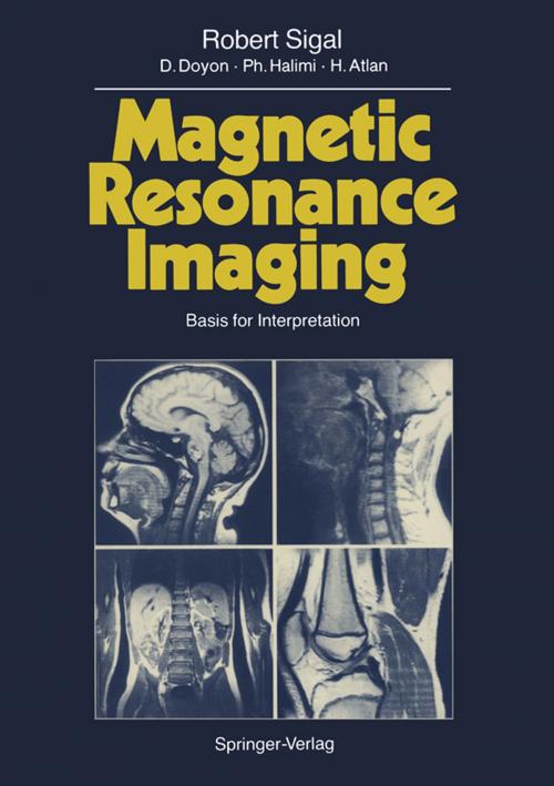
Magnetic Resonance Imaging
Basis for Interpretation
Nonfiction, Health & Well Being, Medical, Specialties, Radiology & Nuclear Medicine, Internal Medicine, Neurology| Author: | Robert Sigal, D. Doyon, P. Halimi, H. Atlan | ISBN: | 9783642730375 |
| Publisher: | Springer Berlin Heidelberg | Publication: | December 6, 2012 |
| Imprint: | Springer | Language: | English |
| Author: | Robert Sigal, D. Doyon, P. Halimi, H. Atlan |
| ISBN: | 9783642730375 |
| Publisher: | Springer Berlin Heidelberg |
| Publication: | December 6, 2012 |
| Imprint: | Springer |
| Language: | English |
Magnetic Resonance Imaging (MRI) is a rapidly evolving technique which is having a significant impact on medical imaging. Only a few years ago, al though Nuclear Magnetic Resonance (NMR) was well known as an important analytical technique in the field of chemical analysis, it was effectively un known in medical circles. Following the initial work of PAUL LAUTERBUR and RAYMOND DAMADIAN in the early 1970s demonstrating that it was possible to use NMR to produce im ages, progress in the medical fields was relatively slow. Recently, however, with the availability of commercial systems, progress has been very rapid, with increasing acceptance of MRI as a basic imaging technique, and the develop ment of exciting new applications. MRI is a relatively complex technique. First, the image depends on many more intrinsic and extrinsic parameters than it does of in techniques like X-ra diography and computed tomography, and secondly, the intrinsic parameters such as T1 and T2 are conceptually complex, involving ideas not usually de scribed in traditional medical imaging courses. In order to produce good MR images efficiently, and to obtain the maximum information from them, it is necessary to appreciate, if not to fully understand, these parameters. Further more, knowledge of how the image is produced helps in appreciating the ori gin of the artifacts sometimes found in MRI due to effects like patient motion and fluid flow.
Magnetic Resonance Imaging (MRI) is a rapidly evolving technique which is having a significant impact on medical imaging. Only a few years ago, al though Nuclear Magnetic Resonance (NMR) was well known as an important analytical technique in the field of chemical analysis, it was effectively un known in medical circles. Following the initial work of PAUL LAUTERBUR and RAYMOND DAMADIAN in the early 1970s demonstrating that it was possible to use NMR to produce im ages, progress in the medical fields was relatively slow. Recently, however, with the availability of commercial systems, progress has been very rapid, with increasing acceptance of MRI as a basic imaging technique, and the develop ment of exciting new applications. MRI is a relatively complex technique. First, the image depends on many more intrinsic and extrinsic parameters than it does of in techniques like X-ra diography and computed tomography, and secondly, the intrinsic parameters such as T1 and T2 are conceptually complex, involving ideas not usually de scribed in traditional medical imaging courses. In order to produce good MR images efficiently, and to obtain the maximum information from them, it is necessary to appreciate, if not to fully understand, these parameters. Further more, knowledge of how the image is produced helps in appreciating the ori gin of the artifacts sometimes found in MRI due to effects like patient motion and fluid flow.
