Transesophageal Echocardiography
Nonfiction, Health & Well Being, Medical, Specialties, Internal Medicine, Cardiology| Author: | Norbert P. de Bruijn, Fiona M. Clements | ISBN: | 9781461320258 |
| Publisher: | Springer US | Publication: | December 6, 2012 |
| Imprint: | Springer | Language: | English |
| Author: | Norbert P. de Bruijn, Fiona M. Clements |
| ISBN: | 9781461320258 |
| Publisher: | Springer US |
| Publication: | December 6, 2012 |
| Imprint: | Springer |
| Language: | English |
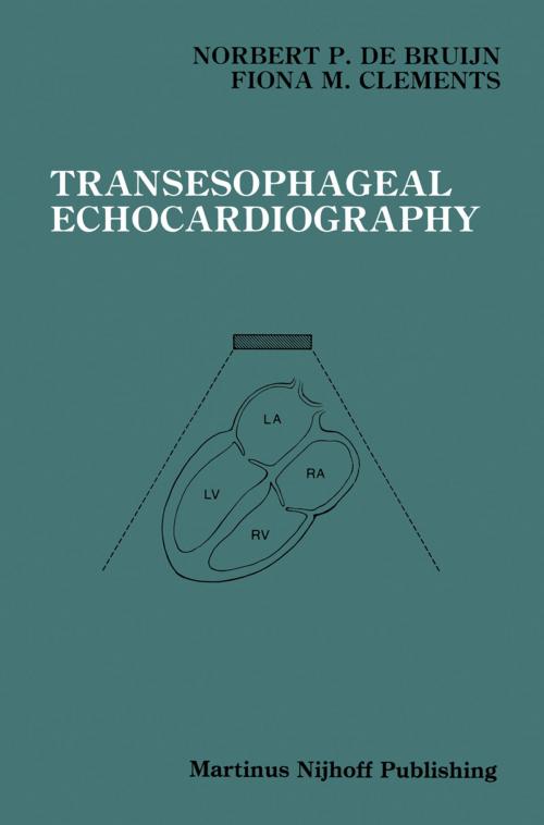
Almost every effort in the care of patients with heart disease begins with some description of disordered physiologic performance or mor phologic anatomy. Since the early work of Edler and Hertz in 1954, echocardiographic methods have grown in importance and reliability for the diagnosis of many cardiac disorders. The placement of a maneuverable transducer on the tip of a modified endoscope is the result of relatively recent technologic advances. The transesophageal approach is now a reality for obtaining new information from ultra-sonic images of a beating human heart. Since images obtained by transesophageal echo cardiography are uniformly of excellent quality, it extends the diagnostic potential of echocardiography to the patient who is difficult to image from the conventional chest wall approach. More importantly, transesophageal echocardiography provides a means to acquire useful information in new situations, such as the operating room. When this imaging modality is brought to patients undergoing surgical procedures, surgeons and anesthesiologists have a ready means for assessing cardiac performance during anesthesia, directing various surgical approaches and immediately evaluating the results of surgical repair. Transesophageal echocardiographic techniques represent a major vi vii advance in the care of patients with cardiovascular disease. Never before has there been a means to acquire such important information about the heart during an operative intervention. New questions are being asked and new answers are at hand.
Almost every effort in the care of patients with heart disease begins with some description of disordered physiologic performance or mor phologic anatomy. Since the early work of Edler and Hertz in 1954, echocardiographic methods have grown in importance and reliability for the diagnosis of many cardiac disorders. The placement of a maneuverable transducer on the tip of a modified endoscope is the result of relatively recent technologic advances. The transesophageal approach is now a reality for obtaining new information from ultra-sonic images of a beating human heart. Since images obtained by transesophageal echo cardiography are uniformly of excellent quality, it extends the diagnostic potential of echocardiography to the patient who is difficult to image from the conventional chest wall approach. More importantly, transesophageal echocardiography provides a means to acquire useful information in new situations, such as the operating room. When this imaging modality is brought to patients undergoing surgical procedures, surgeons and anesthesiologists have a ready means for assessing cardiac performance during anesthesia, directing various surgical approaches and immediately evaluating the results of surgical repair. Transesophageal echocardiographic techniques represent a major vi vii advance in the care of patients with cardiovascular disease. Never before has there been a means to acquire such important information about the heart during an operative intervention. New questions are being asked and new answers are at hand.
