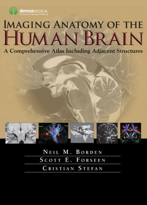
Imaging Anatomy of the Human Brain
A Comprehensive Atlas Including Adjacent Structures
Nonfiction, Health & Well Being, Medical, Reference, Medical Atlases, Specialties, Radiology & Nuclear Medicine, Internal Medicine, Neurology| Author: | Neil M. Borden, MD, Scott E. Forseen, MD, Cristian Stefan, MD | ISBN: | 9781617051258 |
| Publisher: | Springer Publishing Company | Publication: | August 25, 2015 |
| Imprint: | Demos Medical | Language: | English |
| Author: | Neil M. Borden, MD, Scott E. Forseen, MD, Cristian Stefan, MD |
| ISBN: | 9781617051258 |
| Publisher: | Springer Publishing Company |
| Publication: | August 25, 2015 |
| Imprint: | Demos Medical |
| Language: | English |
An Atlas for the 21st Century
The most precise, cutting-edge images of normal cerebral anatomy available today are the centerpiece of this spectacular atlasfor clinicians, trainees, and students in the neurologically-based medical and non-medical specialties. Truly an ìatlas for the 21st century,î this comprehensive visual reference presents a detailed overview of cerebral anatomy acquired through the use of multiple imaging modalities including advanced techniques that allow visualization of structures not possible with conventional MRI or CT. Beautiful color illustrations using 3-D modeling techniques based upon 3D MR volume data sets further enhances understanding of cerebral anatomy and spatial relationships. The anatomy in these color illustrations mirror the black and white anatomic MR images presented in this atlas.
Written by two neuroradiologists and an anatomist who are also prominent educators, along with more than a dozen contributors, the atlasbegins with a brief introduction to the development, organization, and function of the human brain. What follows is more than 1,000 meticulously presented and labelled images acquired with the full complement of standard and advanced modalities currently used to visualize the human brain and adjacent structuresóincluding MRI, CT, diffusion tensor imaging (DTI) with tractography, functional MRI, CTA, CTV, MRA, MRV, conventional 2-D catheter angiography, 3-D rotational catheter angiography, MR spectroscopy, and ultrasound of the neonatal brain. The vast array of data that these modes of imaging provide offers a wider window into the brain and allows the reader a unique way to integrate the complex anatomy presented. Ultimately the improved understanding you can acquire using this atlas can enhance clinical understanding and have a positive impact on patient care. Additionally, various anatomic structures can be viewed from modality to modality and from multiple planes.
This state-of-the-art atlas provides a single source reference, which allows the interested reader ease of use, cross-referencing, and the ability to visualize high-resolution images with detailed labeling. It will serve as an authoritative learning tool in the classroom, and as an invaluable practical resource at the workstation or in the office or clinic.
Key Features:
- Provides detailed views of anatomic structures within and around the human brain utilizing over 1,000 high quality images across a broad range of imaging modalities
- Contains extensively labeled images of all regions of the brain and adjacent areas that can be compared and contrasted across modalities
- Includes specially created color illustrations using computer 3-D modeling techniques to aid in identifying structures and understanding relationships
- Goes beyond a typical brain atlas with detailed imaging of skull base, calvaria, facial skeleton, temporal bones, paranasal sinuses, and orbits
- Serves as an authoritative learning tool for students and trainees and practical reference for clinicians in multiple specialties


