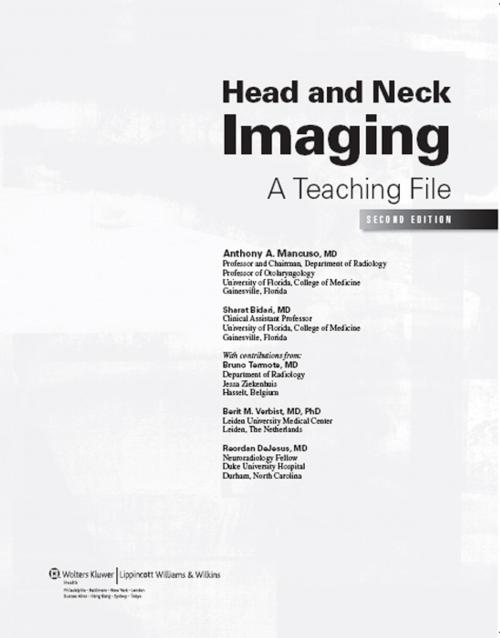
Head and Neck Imaging
A Teaching File
Nonfiction, Health & Well Being, Medical, Specialties, Radiology & Nuclear Medicine| Author: | Anthony A. Mancuso, Sharat Bidari, Bruno Termote, Berit M. Verbist, Reordan DeJesus | ISBN: | 9781451154313 |
| Publisher: | Wolters Kluwer Health | Publication: | January 5, 2012 |
| Imprint: | LWW | Language: | English |
| Author: | Anthony A. Mancuso, Sharat Bidari, Bruno Termote, Berit M. Verbist, Reordan DeJesus |
| ISBN: | 9781451154313 |
| Publisher: | Wolters Kluwer Health |
| Publication: | January 5, 2012 |
| Imprint: | LWW |
| Language: | English |
Teaching Files are one of the hallmarks of radiology education, providing the kind of personal consultation with experts normally found only in the setting of a teaching hospital. This teaching file contains 164 cases, covering all areas of head and neck imaging. A consistent format is used to present each case. Chapters are organized by anatomical region. Included are cases on the temporal bone, skull base, eye and orbit, sinuses and nasal cavity, neck, trachea, salivary glands, hypopharynx and cervical esophagus, oropharynx and larynx. The book can be used both as an independent study and review tool for board-exam preparation and as a companion to Dr. Mancuso's majesterial, two-volume text, "Head and Neck Radiology" (2010), chapters from which are referenced following each case for further reading. The cases follow a standard format popular with residents and fellows: clinical presentation, questions, findings, differential diagnosis, diagnosis, discussion, questions for further thought, reporting responsibilities, and a section on what the treating physician needs to know. Cases conclude with answers to the questions. This format emphasizes critical thinking within a clinical context, to build medical decision-making skills. A password-protected companion website, free to purchasers of the book, includes all of the cases for online review.
Teaching Files are one of the hallmarks of radiology education, providing the kind of personal consultation with experts normally found only in the setting of a teaching hospital. This teaching file contains 164 cases, covering all areas of head and neck imaging. A consistent format is used to present each case. Chapters are organized by anatomical region. Included are cases on the temporal bone, skull base, eye and orbit, sinuses and nasal cavity, neck, trachea, salivary glands, hypopharynx and cervical esophagus, oropharynx and larynx. The book can be used both as an independent study and review tool for board-exam preparation and as a companion to Dr. Mancuso's majesterial, two-volume text, "Head and Neck Radiology" (2010), chapters from which are referenced following each case for further reading. The cases follow a standard format popular with residents and fellows: clinical presentation, questions, findings, differential diagnosis, diagnosis, discussion, questions for further thought, reporting responsibilities, and a section on what the treating physician needs to know. Cases conclude with answers to the questions. This format emphasizes critical thinking within a clinical context, to build medical decision-making skills. A password-protected companion website, free to purchasers of the book, includes all of the cases for online review.
