CT Densitometry in Osteoporosis
The impact on management of the patient
Nonfiction, Health & Well Being, Medical, Medical Science, Diagnostic Imaging, Specialties, Orthopedics| Author: | L.E. Lampmann, S.A. Duursma, J.H.J. Ruys | ISBN: | 9789400956728 |
| Publisher: | Springer Netherlands | Publication: | December 6, 2012 |
| Imprint: | Springer | Language: | English |
| Author: | L.E. Lampmann, S.A. Duursma, J.H.J. Ruys |
| ISBN: | 9789400956728 |
| Publisher: | Springer Netherlands |
| Publication: | December 6, 2012 |
| Imprint: | Springer |
| Language: | English |
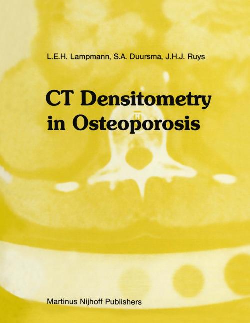
In 1977 a Philips Tomoscan 200, second generation, whole body CT scanner was installed at the Department of Radiodiagnosis of the University Hospital of Utrecht (The Netherlands) and its new possibilities concerning the measurements of bone mineral content (BMC) had been considered. As a result of the close cooperation between the Clinical Research Group for Bone Metabolism and the Department of Radiodiagnosis of the University Hospital of Utrecht a new project was started. The aim of a pilot study was to investigate the application of CT scanning in BMC determination in comparison with existing parameters such as histovolumetric measurements in transiliac bone biopsy specimens, morphometric measurements in hand X-ray films and other methods. In 1979 a Philips Tomoscan 300, third generation CT scanner became available. With this new scanner many problems of the Tomoscan 200 seemed to be solved. An examination protocol was designed with a standardized method for CT mea surements and a follow-up study was started. On account of the availability and the participation of a number of patients it became possible to realize this study. The results made it possible to draw conclusions concerning CT densitometry, with an impact on the management of the osteoporotic patient. A new dimension is added to the diagnostic procedures concerning osteoporosis and to the methods for measuring the effect of therapeutic regimes. Our aim is to offer the reader insight in the possibilities and limitations of this technique, compared with other parameters in BMC determination.
In 1977 a Philips Tomoscan 200, second generation, whole body CT scanner was installed at the Department of Radiodiagnosis of the University Hospital of Utrecht (The Netherlands) and its new possibilities concerning the measurements of bone mineral content (BMC) had been considered. As a result of the close cooperation between the Clinical Research Group for Bone Metabolism and the Department of Radiodiagnosis of the University Hospital of Utrecht a new project was started. The aim of a pilot study was to investigate the application of CT scanning in BMC determination in comparison with existing parameters such as histovolumetric measurements in transiliac bone biopsy specimens, morphometric measurements in hand X-ray films and other methods. In 1979 a Philips Tomoscan 300, third generation CT scanner became available. With this new scanner many problems of the Tomoscan 200 seemed to be solved. An examination protocol was designed with a standardized method for CT mea surements and a follow-up study was started. On account of the availability and the participation of a number of patients it became possible to realize this study. The results made it possible to draw conclusions concerning CT densitometry, with an impact on the management of the osteoporotic patient. A new dimension is added to the diagnostic procedures concerning osteoporosis and to the methods for measuring the effect of therapeutic regimes. Our aim is to offer the reader insight in the possibilities and limitations of this technique, compared with other parameters in BMC determination.
