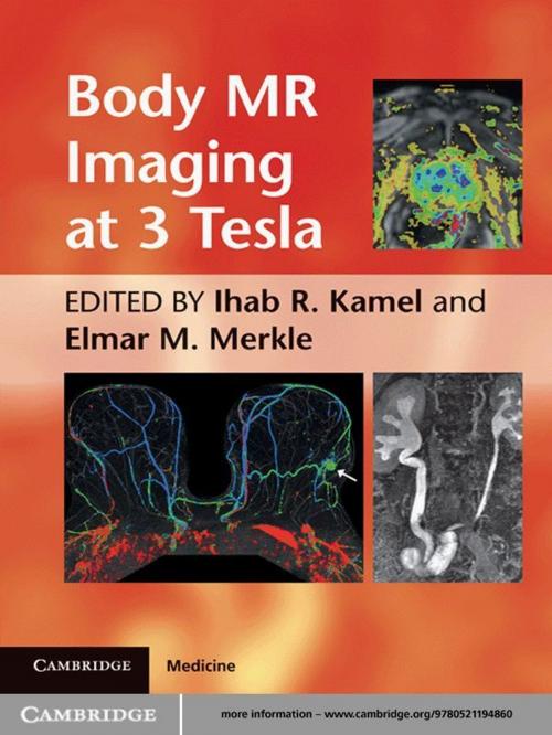
Body MR Imaging at 3 Tesla
Nonfiction, Health & Well Being, Medical, Medical Science, Diagnostic Imaging, Specialties, Radiology & Nuclear Medicine| Author: | ISBN: | 9781139088664 | |
| Publisher: | Cambridge University Press | Publication: | August 4, 2011 |
| Imprint: | Cambridge University Press | Language: | English |
| Author: | |
| ISBN: | 9781139088664 |
| Publisher: | Cambridge University Press |
| Publication: | August 4, 2011 |
| Imprint: | Cambridge University Press |
| Language: | English |
Body MR Imaging at 3.0 Tesla is a practical text enabling radiologists to maximise the benefits of high field 3T MR systems in a range of body applications. It explains the physical principles of MR imaging using 3T magnets, and the differences between 1.5T and 3T when applied extracranially. The book's organ-based approach focuses on optimized techniques, providing recommended protocols for the main vendors of 3T MRI systems. All major thoracic and abdominal organs are covered, including breast, heart, liver, pancreas, the GI tract, kidneys, prostate and female pelvic organs. Abdominal and pelvic MR angiography and MRCP are also discussed. Protocol optimization, appearance of artifacts and novel applications using 3T are emphasized. Written and edited by experts in the field, Body MR Imaging at 3.0 Tesla guides radiologists in optimizing imaging protocols for 3T MR systems, reducing artifacts and identifying the advantages of using 3T in body applications.
Body MR Imaging at 3.0 Tesla is a practical text enabling radiologists to maximise the benefits of high field 3T MR systems in a range of body applications. It explains the physical principles of MR imaging using 3T magnets, and the differences between 1.5T and 3T when applied extracranially. The book's organ-based approach focuses on optimized techniques, providing recommended protocols for the main vendors of 3T MRI systems. All major thoracic and abdominal organs are covered, including breast, heart, liver, pancreas, the GI tract, kidneys, prostate and female pelvic organs. Abdominal and pelvic MR angiography and MRCP are also discussed. Protocol optimization, appearance of artifacts and novel applications using 3T are emphasized. Written and edited by experts in the field, Body MR Imaging at 3.0 Tesla guides radiologists in optimizing imaging protocols for 3T MR systems, reducing artifacts and identifying the advantages of using 3T in body applications.


